Table of Contents
Understanding Sialendoscopy: A Breakthrough in Salivary Gland Treatment

Sialendoscopy is an innovative minimally invasive procedure used to diagnose and treat various salivary gland disorders. This technique has been a game-changer in the field of otolaryngology, particularly for conditions such as sialolithiasis (the formation of salivary stones) and chronic sialadenitis (inflammation of the salivary glands). Sialendoscopy allows for direct visualization of the salivary ducts using a micro-endoscope, enabling surgeons to remove obstructions, flush out debris, and dilate strictures without the need for extensive surgical intervention.
Historically, treatments for salivary gland disorders often involved more invasive methods, such as gland excision, which could result in significant complications, including nerve damage, scarring, and prolonged recovery times. The advent of sialendoscopy, which employs tools as delicate as the tip of a ballpoint pen, has transformed patient management by reducing these risks and improving recovery outcomes (Puscas, 2024).
The Anatomy of Salivary Glands and Their Role in Health

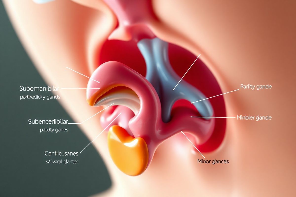
The human salivary glands consist of three major pairs: the parotid, submandibular, and sublingual glands, along with numerous minor salivary glands throughout the oral cavity. These glands are critical components of the digestive system, responsible for producing saliva, which aids in moistening food, facilitating swallowing, and initiating digestion. Saliva also plays a crucial role in oral health, helping to prevent tooth decay by neutralizing acids and providing antimicrobial properties (NIDCR, 2024).
Salivary Gland Locations and Functions
| Gland Type | Location | Function |
|---|---|---|
| Parotid | In front of each ear | Produces a watery saliva rich in enzymes |
| Submandibular | Beneath the jaw | Produces a mixed saliva (serous and mucous) |
| Sublingual | Under the tongue | Primarily produces mucous saliva |
| Minor Salivary Glands | Throughout the mouth | Contribute to overall saliva production |
Understanding the anatomy of these glands is vital for diagnosing and treating disorders effectively. Conditions affecting the salivary glands can lead to significant discomfort and complications if not addressed promptly.
Common Salivary Gland Disorders: Symptoms and Causes
Salivary gland disorders encompass a range of conditions, with the most common being sialolithiasis and sialadenitis. These disorders can lead to a variety of symptoms, including:
- Pain and swelling in the affected gland, particularly when eating.
- Dry mouth (xerostomia), often resulting from insufficient saliva production.
- Difficulty swallowing or speaking due to gland enlargement or blockage.
- Foul taste or bad breath, which may indicate infection or stagnant saliva.
Causes of Salivary Gland Disorders
The underlying causes of salivary gland disorders can include:
- Sialolithiasis (salivary stones): These are calcified deposits that obstruct the salivary ducts, leading to swelling and pain (Healthline, 2024).
- Infections: Bacterial infections commonly occur in glands that are obstructed or have low saliva flow. Conditions like Sjögren’s syndrome, which is an autoimmune disorder, can lead to chronic dry mouth and recurrent infections (Merck Manual, 2024).
- tumors: Both benign and malignant tumors can develop in the salivary glands, causing noticeable swelling and other symptoms (NIDCR, 2024).
- Radiation therapy: Treatments for head and neck cancers can damage salivary glands, leading to long-term complications such as dry mouth (Sridharan, 2024).
The Sialendoscopy Procedure: What to Expect
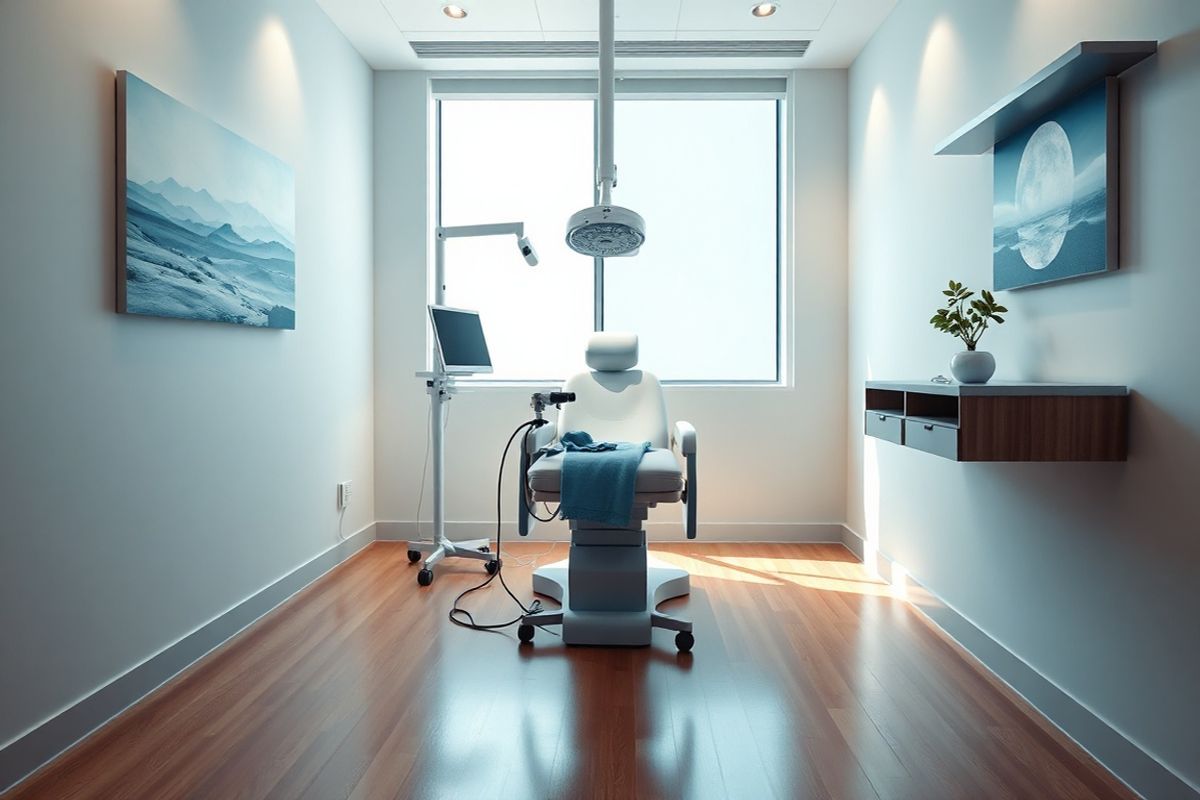
The sialendoscopy procedure is typically performed in an outpatient setting under local or general anesthesia. Patients can expect the following steps during the procedure:
- Preparation: Patients may undergo imaging studies, such as ultrasound or CT scans, to identify stones or obstructions (Hopkins Medicine, 2024).
- Anesthesia: Local or general anesthesia is administered to ensure comfort during the procedure.
- Insertion of the Endoscope: A micro-endoscope is gently inserted into the duct of the affected gland. The duct may be dilated if it is too tight.
- Removal of Obstructions: The physician can use small instruments to remove stones or debris and irrigate the gland with saline to flush out any remaining materials.
- Post-Procedure Care: After the procedure, patients may experience some soreness but typically recover quickly, returning to normal activities within a few days (Duke Health, 2024).
Benefits of Sialendoscopy
The benefits of sialendoscopy over traditional surgical methods include:
- Minimally invasive: Reduced risk of complications and shorter recovery times.
- Preservation of gland function: Unlike gland removal, sialendoscopy aims to maintain the gland’s ability to produce saliva.
- Immediate symptom relief: Many patients experience rapid improvement in symptoms following the procedure.
Evaluating the Benefits and Risks of Sialendoscopy
Like any medical procedure, sialendoscopy has its benefits and risks.
Benefits
- Minimized Recovery Time: Patients often return home the same day and experience shorter recovery periods compared to traditional surgery.
- Reduced Complications: The minimally invasive nature of the procedure lowers the risk of complications such as nerve damage, scarring, and longer-term dysfunction of the salivary glands.
- High Success Rate: Sialendoscopy has shown a success rate exceeding 80% for relieving symptoms associated with salivary stones (Sridharan, 2024).
Risks
- Infection: While rare, there is a possibility of infection following the procedure.
- Strictures: There is a risk of scarring or strictures developing in the duct.
- Perforation: In rare cases, the duct may be inadvertently perforated during the procedure.
Conclusion
Sialendoscopy represents a significant advancement in the treatment of salivary gland disorders, offering a less invasive option that preserves gland function and provides quick relief from painful symptoms. As with any medical procedure, patients should discuss the risks and benefits with their healthcare provider to determine the best course of action for their specific condition.
Frequently Asked Questions (FAQs)
What is sialendoscopy?
Sialendoscopy is a minimally invasive procedure used to diagnose and treat salivary gland disorders, particularly those caused by blockages like salivary stones.
How long does the sialendoscopy procedure take?
The procedure typically takes about 90 minutes to complete.
What can I expect after the procedure?
Patients may experience some soreness, but most can return to normal activities within a few days.
Are there any risks associated with sialendoscopy?
Yes, potential risks include infection, duct strictures, and perforation, although these complications are rare.
How can I prevent salivary gland disorders?
Staying hydrated, maintaining good oral hygiene, and managing underlying medical conditions can help reduce the risk of developing salivary gland disorders.
References
- Puscas. (2024). Sialendoscopy Removes Salivary Stones, Spares Glands, Speeds Recovery. Retrieved from https://www.dukehealth.org/blog/sialendoscopy-removes-salivary-stones-spares-glands-speeds-recovery
- NIDCR. (2024). Saliva & Salivary Gland Disorders. Retrieved from https://www.nidcr.nih.gov/health-info/saliva-salivary-gland-disorders
- Healthline. (2024). Salivary Gland Disorders: Causes, Symptoms, and Diagnosis. Retrieved from https://www.healthline.com/health/salivary-gland-disorders
- Merck Manual. (2024). Salivary Gland Disorders. Retrieved from https://www.merckmanuals.com/home/ear-nose-and-throat-disorders/mouth-and-throat-disorders/salivary-gland-disorders
- Sridharan, S. (2024). Sparing Surgery and the Role of Sialendoscopy. Retrieved from https://eyeandear.org/2024/06/salivary-gland-sparing-surgery-and-the-role-of-sialendoscopy
- Hopkins Medicine. (2024). Sialendoscopy. Retrieved from https://www.hopkinsmedicine.org/health/treatment-tests-and-therapies/sialendoscopy


