Table of Contents
Understanding Clotting Factors: The Building Blocks of Hemostasis

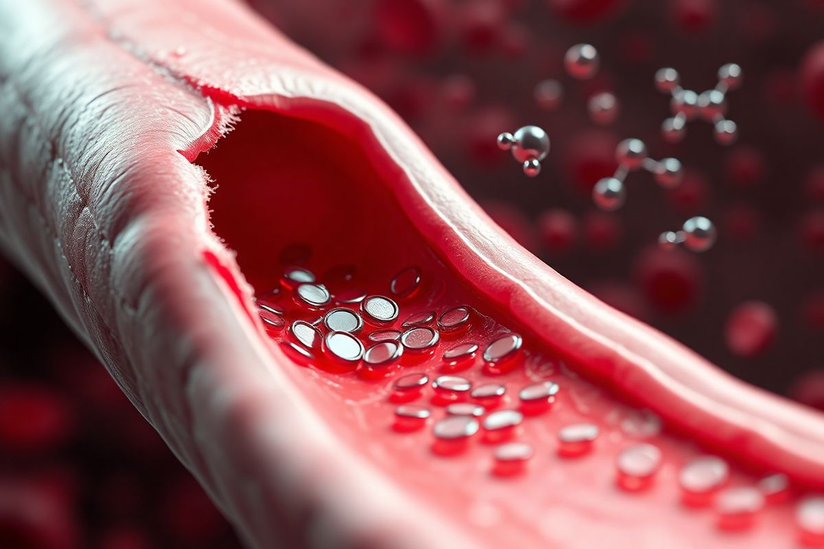
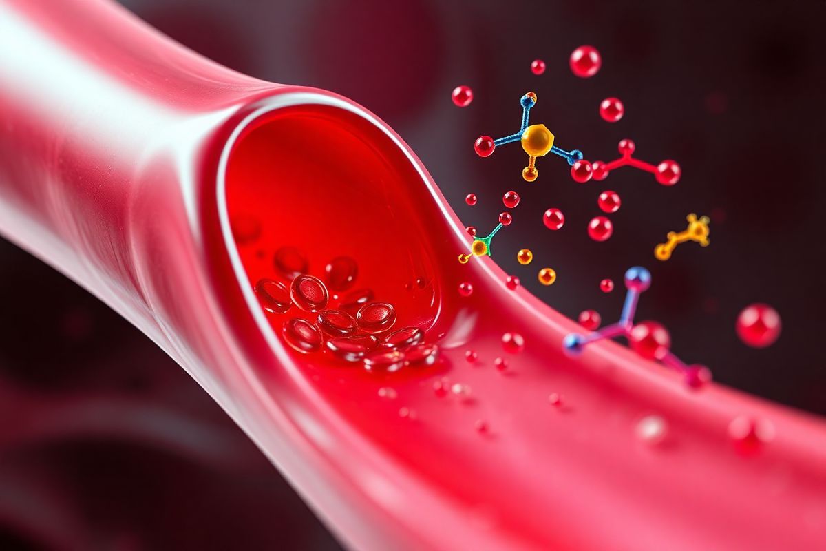
Hemostasis, the process that prevents and stops bleeding, is crucial for maintaining the integrity of the circulatory system. This complex process involves a delicate balance between coagulation and anticoagulation mechanisms. At the heart of hemostasis are blood clotting factors, a series of proteins that play vital roles in the formation of blood clots. These factors are primarily produced in the liver and circulate in an inactive form until they are activated in response to vascular injury.
Clotting factors are typically denoted by Roman numerals (I through XIII), with each factor playing a specific role in the coagulation cascade. For instance, Factor I, known as fibrinogen, is converted into fibrin, which forms the mesh-like structure of a clot. Factors II (prothrombin), III (tissue factor), and X (Stuart-Prower factor) are also pivotal, as they facilitate the conversion of prothrombin to thrombin, a key enzyme that catalyzes fibrin formation. The interplay among these factors ensures rapid clot formation to prevent excessive bleeding following an injury (National Heart, Lung, and Blood Institute, 2022).
The Intricate Blood Clotting Process: From Injury to Healing

The blood clotting process begins immediately after an injury to a blood vessel. When a vessel is damaged, the endothelial cells lining the blood vessels release chemical signals that prompt the constriction of the vessel to minimize blood loss. Platelets, the tiny cells responsible for clot formation, are activated and migrate to the site of injury. They adhere to the exposed collagen fibers and each other, forming a temporary “platelet plug” (What Causes Blood to Coagulate?, 2022).
Once the platelet plug is formed, a cascade of coagulation factors is activated, leading to the conversion of fibrinogen into fibrin. This transformation results in a stable blood clot, which effectively seals the wound and prevents further bleeding. The clotting process can be summarized in several stages:
- Vasoconstriction: The injured blood vessel narrows to reduce blood flow.
- Platelet Activation: Platelets adhere to the injury site, becoming activated and aggregating to form a plug.
- Coagulation Cascade: Activated platelets release signaling molecules that activate clotting factors, leading to fibrin formation.
- Clot Retraction and Repair: The clot contracts to approximate the edges of the wound, allowing tissue repair mechanisms to initiate (Blood Clotting Disorders - How Does Blood Clot?, 2022).
This entire process is tightly regulated. Any dysfunction in the clotting factors or platelets can lead to bleeding disorders or excessive clotting.
von Willebrand Factor: A Key Player in the Coagulation Cascade
Among the various clotting factors, von Willebrand factor (vWF) stands out as a critical component in the coagulation cascade. vWF is a large glycoprotein synthesized by endothelial cells and megakaryocytes, and it plays a pivotal role in hemostasis by facilitating platelet adhesion to sites of vascular injury.
When a blood vessel is damaged, vWF binds to exposed collagen and acts as a bridge between the collagen and platelets. This interaction is essential for the formation of the initial platelet plug, particularly under conditions of high shear stress, such as in arteries. In addition to its adhesive properties, vWF also serves as a carrier protein for Factor VIII, another crucial clotting factor that is involved in the intrinsic pathway of coagulation. The protection and stabilization of Factor VIII by vWF are vital for effective clotting, as deficiencies in vWF can lead to hemophilia A (Explainer: What Are Blood Clotting Factors?, 2024).
vWF exists in several multimers, which vary in size and functionality. Larger vWF multimers are more effective in promoting platelet aggregation than smaller ones. The regulation of vWF levels and its multimeric structure is essential for maintaining normal hemostatic function. Conditions such as von Willebrand disease, the most common inherited bleeding disorder, result from qualitative or quantitative deficiencies in vWF, leading to impaired platelet adhesion and increased bleeding risk (Senst et al., 2021).
How Clotting Factors Work Together to Form a Stable Blood Clot
The formation of a stable blood clot is a sophisticated interplay between various clotting factors, platelets, and the vascular system. The coagulation cascade is generally divided into three pathways: the intrinsic pathway, the extrinsic pathway, and the common pathway.
- Intrinsic Pathway: Triggered by damage to the blood vessel, this pathway involves several clotting factors (Factors XII, XI, IX, and VIII). It culminates in the activation of Factor X.
- Extrinsic Pathway: This pathway is activated by tissue factor (Factor III), which is exposed upon vessel injury. It rapidly leads to the activation of Factor X.
- Common Pathway: Both intrinsic and extrinsic pathways converge here, leading to the conversion of prothrombin (Factor II) to thrombin (Factor IIa), which then converts fibrinogen into fibrin strands, forming a mesh that solidifies the platelet plug.
The coordinated activation of these factors leads to a rapid and effective response to vascular injury. The clot is then stabilized by the action of Factor XIII, which cross-links fibrin, enhancing the structural integrity of the clot.
| Pathway | Key Factors Involved |
|---|---|
| Intrinsic | Factors XII, XI, IX, VIII |
| Extrinsic | Factor III (tissue factor) |
| Common | Factors II (prothrombin), X, XIII |
The balance between clot formation and breakdown is equally important. Fibrinolysis, the process by which clots are dissolved, is initiated by the conversion of plasminogen to plasmin, which breaks down fibrin into soluble fragments. This process is crucial for maintaining homeostasis and preventing excessive clotting (How Blood Clots, 2022).
The Consequences of Clotting Factor Deficiencies: Risks and Disorders
Deficiencies in any of the clotting factors can lead to serious health risks and bleeding disorders. Two well-known disorders related to clotting factor deficiencies are hemophilia and von Willebrand disease.
-
Hemophilia: This genetic disorder is characterized by deficiencies in specific clotting factors. Hemophilia A is due to a deficiency of Factor VIII, while Hemophilia B results from a deficiency in Factor IX. Patients with hemophilia experience prolonged bleeding, especially after injuries or surgeries, and may have spontaneous bleeding episodes.
-
Von Willebrand Disease: This is caused by a deficiency or dysfunction of von Willebrand factor. Patients may experience symptoms such as frequent nosebleeds, easy bruising, and heavy menstrual bleeding. Unlike hemophilia, von Willebrand disease is more common and often affects both men and women (National Heart, Lung, and Blood Institute, 2022).
Other clotting disorders can include thrombosis-related conditions, where excessive clotting occurs, leading to complications such as deep vein thrombosis (DVT), pulmonary embolism, or stroke. Understanding and diagnosing these conditions often involves various coagulation factor tests that assess the levels and functionality of specific clotting factors in the blood.
| Disorder | Key Characteristics |
|---|---|
| Hemophilia A | Deficiency of Factor VIII, prolonged bleeding |
| Hemophilia B | Deficiency of Factor IX, prolonged bleeding |
| Von Willebrand Disease | Deficiency or dysfunction of vWF, easy bruising |
FAQ
What is von Willebrand factor? von Willebrand factor is a glycoprotein that plays a critical role in blood clotting by promoting platelet adhesion to damaged blood vessels.
What are the symptoms of von Willebrand disease? Symptoms can include frequent nosebleeds, easy bruising, heavy or prolonged menstrual bleeding, and excessive bleeding after injury or surgery.
How is von Willebrand disease diagnosed? Diagnosis typically involves blood tests to measure the level and functionality of von Willebrand factor and other clotting factors.
Can von Willebrand disease be treated? Yes, treatment may include medications that increase the levels of von Willebrand factor in the blood or clotting factor replacement therapy.
What are the risks of clotting factor deficiencies? Risks include excessive bleeding, spontaneous bleeding episodes, and complications such as internal bleeding, which can be life-threatening.
References
- National Heart, Lung, and Blood Institute. (2022). Blood Clotting Disorders - How Does Blood Clot? Retrieved from https://www.nhlbi.nih.gov/health/clotting-disorders/how-blood-clots
- What Causes Blood to Coagulate? (2022). Retrieved from https://www.bleedingdisorders.com/about/how-blood-clots-coagulation
- Explainer: What Are Blood Clotting Factors? (2024). Retrieved from https://www.csl.com/we-are-csl/vita-original-stories/2024/explainer-what-are-blood-clotting-factors
- How Blood Clots. (2022)
- Senst, B., Tadi, P., Goyal, A., et al. (2021). Effect of endothelial responses on sepsis-associated organ dysfunction. Retrieved from https://pubmed.ncbi.nlm.nih.gov/11649038/


